E-ISSN:2349-4166
P-ISSN:2349-4158

Research Article

Desensitizing

Public Health Review - International Journal of Public Health Research

2019 Volume 6 Number 4 July-August
Publisherwww.medresearch.in

A comparative evaluation of the effect of four desensitizing agents (strontium chloride, potassium nitrate, pro-argin, bioactive glass) on dentinal occlusion: An ex-vivo SEM study

Hongal S.1*, Arjun Torwane N.2, Hiremath V.3, Jain M.4
DOI: https://doi.org/10.17511/ijphr.2019.i4.03

1* Sudheer Hongal, Professor & Head, Department of Public Health Dentistry, HKDET’S Dental College and Hospital, Humnabad, Karnataka, India.

2 Nilesh Arjun Torwane, Reader, Department of Public Health Dentistry, Peoples Dental Academy, Bhopal, Madhya Pradesh, India.

3 Vinay Hiremath, Principal & Professor, Department of Oral Pathology, HKDET’S Dental College and Hospital, Humnabad, Karnataka, India.

4 Manish Jain, Professor, SMBT Institute of Dental Sciences and Research, Dhamangaon, Dhamangaon, India.

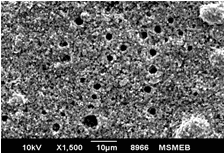
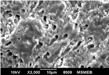
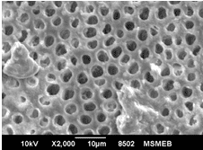
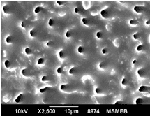
Aim: The aim of this study was to compare and evaluate the effect of four desensitizing agents (Strontium Chloride, Potassium Nitrate, Pro-Argin, Bioactive Glass) on dentinal occlusion. Materials and Methods: Forty freshly extracted mandibular molars were selected. Dentine discs measuring 6x6x3mm were cut off from cervical 1/3rd of molars. The occlusal surface of each dentine disc was abraded with silicon carbide paper for 1 minute and then immersed in 17% Ethylenediaminetetra-acetic acid for 5 minutes. Results: Data were statistically analyzed by one-way Analysis of Variance (ANOVA) and Tukey’s test. The P value for each group was <0.001 showing that highly significant difference existed among the groups. The Bioactive Glass showed maximum amount of dentinal tubule occlusion among all the groups. The least amount of dentinal tubule occlusion was observed in potassium nitrate. Conclusion: It was concluded that tooth brushing with dentifrice containing bioactive glass showed maximum amount of dentine tubule occlusion. Clinical Relevance: Dentinal hypersensitivity is a significant clinical problem which can cause considerable concern for patients. Appropriate and combine use of various clinically effective both synthetic and natural desensitizing agents may considerably help to manage the problem.

Keywords: Dentinal hypersensitivity, Dentinal occlusion, Desensitizing agent

Corresponding Author How to Cite this Article To Browse
Sudheer Hongal, Professor & Head, Department of Public Health Dentistry, HKDET’S Dental College and Hospital, Humnabad, Karnataka, India.
Email:
Hongal S, Torwane NA, Hiremath V, Jain M. A comparative evaluation of the effect of four desensitizing agents (strontium chloride, potassium nitrate, pro-argin, bioactive glass) on dentinal occlusion: An ex-vivo SEM study. Public Health Rev Int J Public Health Res. 2019;6(4):147-153.
Available From
https://publichealth.medresearch.in/index.php/ijphr/article/view/113

Manuscript Received Review Round 1 Review Round 2 Review Round 3 Accepted
2019-07-05 2019-07-15 2019-07-20 2019-07-24
Conflict of Interest Funding Ethical Approval Plagiarism X-checker Note
No Nil Yes 8%

© 2019 by Sudheer Hongal, Nilesh Arjun Torwane, Vinay Hiremath, Manish Jain and Published by Siddharth Health Research and Social Welfare Society. This is an Open Access article licensed under a Creative Commons Attribution 4.0 International License https://creativecommons.org/licenses/by/4.0/ unported [CC BY 4.0].

Introduction

Dentinal hypersensitivity is one of the most commonly encountered clinical problems. According to Addy et al. dentine hypersensitivity is characterized by “Pain derived from exposed dentine in response to chemical, thermal, tactile, or osmotic stimuli which cannot be explained as arising from any other dental defect or pathology’’[1]. Dentinal hypersensitivity is typically experienced by young to middle-aged adults, with incidence of 4 to 74% of the population [2, 3]. Several theories have been suggested to explain the mechanism of dentinal hypersensitivity like odontoblastic transduction theory and neural theory. By far the most widely accepted theory is the hydrodynamic theory proposed by Brannstrom et al. This theory postulates that fluids within the dentinal tubules are disturbed either by thermal, physical, chemical or osmotic changes and these fluid changes or movements stimulate a baroreceptor which leads to neural discharge [4, 5]. Various chemical agents have been used for dentine hypersensitivity treatment. The goal of such therapies is to prevent the pain signal from being triggered [6]. Various chemical compounds have been used such as aluminium lactate, sodium fluoride, kaolin and glycerine paste, strontium fluoride, ferric and aluminium oxalate, sodium monoflourophosphate, stannous fluoride, potassium nitrate, potassium oxalate, cyanoacrylate, resins, hydroxyapatite and bioactive glass [7]. Dentifrices containing 10% strontium chloride have been widely used as desensitizing agents. It has been proposed that the ions occlude dentinal tubules by binding to the tooth substance.6 Strontium ions penetrate dentine deeply and replace calcium, resulting in recrystallization in the form of a strontium apatite complex [8]. Potassium salts block neural transmission at the pulp and depolarize the nerve around the odontoblasts. Potassium nitrate has been incorporated into both toothpastes and mouth rinses for use as a treatment for dental hypersensitivity. Kim reported for the first time that the potassium ion is the active portion of potassium nitrate [6]. Pro-Argin, developed by Kleinberg et al., consists of 8% arginine with bicarbonate and calcium carbonate. The rationale behind this composition is that arginine, an amino acid is positively charged at physiological pH, bicarbonate is a pH buffer, and calcium carbonate is a source of calcium. This composition helps in maintaining the alkaline pH of saliva and hence favours tubular

occlusion by forming a glycoprotein on the surface [9]. Bioactive glass is an inorganic amorphous calcium sodium phosphosilicate, originally developed as bone regenerative material. These materials are reactive when exposed to body fluids, and deposit hydroxycarbonate apatite, as a mineral in the enamel and dentine. When incorporated into a dentifrice, particles are deposited onto the dentine surface to mechanically occlude dentinal tubules [10]. Therefore, the aim of this study was to evaluate and compare the ability of four desensitizing agents – Strontium Chloride, Potassium Nitrate, Pro-Argin and Bioactive Glass on dentinal tubule occlusion using a scanning electron microscope.

Materials and Methods

In-Vitro Study Setting: Forty freshly extracted mandibular molars were selected for the study. Teeth were immersed in 5% sodium hypochlorite (Dentpro, India) for 2 hours and then stored in 10% formalin (Dentpro, India) for complete disinfection. The calculi and soft tissue remnants on the external surface were removed with a periodontal curette.

Procedure : The teeth were sectioned such that the first cut was made perpendicular to the long axis of the tooth above the cemento-enamel junction by means of a diamond disc and then the occlusal surfaces of the teeth were removed with a second cut parallel to the first, to remove all the coronal enamel to expose the flat dentine surfaces to obtain 3mm thick dentinal discs from cervical 1/3rd of each molar [11].

The dentine discs were cut off measuring 6x6x3 mm. The prepared tooth specimens were then mounted on an acrylic block. After preparation of the specimens, the occlusal surface of each dentine disc was abraded with 400 and 1000 grit silicon carbide paper for 1 minute to create a standard smear layer. The smear layer was subsequently removed by dipping the dentine disc into 17% Ethylenediaminetetra-acetic acid solution (Canalarge, Ammdent, India) for 5 minutes. The demineralized dentine discs were then rinsed by distilled water and kept wet.

Random allocation: The specimens were then randomly distributed into 4 groups each containing 10 specimens.

Group 1: Specimens were brushed with Strontium Chloride containing dentifrice Thermoseal (ICPA Health Products Ltd., India) for 2 min for 7 days.


Group 2: Specimens were brushed with Potassium Nitrate containing dentifrice Sensodyne (Glaxo Smith Kline Asia Pvt. Ltd., India) for 2 min for 7 days.

Group 3: Specimens were brushed with Pro-Argine containing dentifrice Colgate Sensitive Pro Relief (Colgate-Palmolive Ltd., India) for 2 min for 7 days.

Group 4: Specimens were brushed with Bioactive Glass containing dentifrice Shy NM (Group Pharma-ceuticals Ltd., India) for 2 min for 7 days.

Each specimen was brushed with undiluted tooth paste (1 gm) with colgateactibrush which is battery powered with soft round bristle and oscillation speed of 8000/min at an inclination of 900. After each brushing session, specimens were rinsed with distilled water and stored in artificial saliva for 7 days [11]. Specimens obtained post-treatment were washed with distilled water, and then these were sputter-coated with 25 nm thin layer of gold.

Scanning electron microscope readings: Photomicro-graphs of the dentine surface were obtained using Scanning Electron Microscope (ZEISS, EVO 50, Germany) at 1000X, 2000X and 5000X magnification.

Percentage of occluded tubules was obtained by dividing the total number of occluded tubules by the

total number of tubules in each photomicrograph. This result was multiplied by 100, to obtain the percentage of occluded tubules [11]. The specimens used for SEM analysis were examined by Energy-dispersive X-ray (EDX) microanalysis to determine the presence of chemical elements in deposits found next to the dentinal tubules of each specimen. The spectrum was obtained at 20kV, with a spot size of 5 nm and a counting time of 300 seconds. EDX analysis provides a qualitative information [12].

Statistical Analysis: Results were statistically analyzed by one-way Analysis of Variance (ANOVA) and Tukey’s test. SPSS 14 software was used for all the analysis. A P-value of <0.05 was considered statistically significant.

Results

The mean values are given in Table 1. Two-way ANOVA showed that all groups presented a significant tubule occlusion after 7 days for the factor time (P < 0.001). Comparison of four groups using post-hoc Tukey’s test in Table 3 showed that Bioactive Glass showed maximum amount of dentinal tubule occlusion among all the groups. The least amount of dentinal tubule occlusion was observed in potassium nitrate.


Table-1: Comparison of mean values of the test groups by using two way ANOVA

Test groups Statistical significance
Strontium Chloride containing dentifrice (Thermoseal) Potassium Nitrate containing dentifrice (Sensodyne) Pro-Argine containing dentifrice (Colgate Sensitive Pro Relief) Bioactive glass containing dentifrice (Shy NM)
Mean S.D. Mean S.D. Mean S.D. Mean S.D.
50.54 6.74 20.81 3.05 40.48 2.31 85.49 6.05 P<0.001

S.D.: Standard Deviation

*Statistical Significance: P<0.005

Table-2: Comparison of mean values between the test groups by using post-hoc Tukey’s test

Test Groups Mean Difference Statistical Significance
Thermoseal vs Sensodyne 29.72 P<0.001*
Thermosealvs Colgate Sensitive Pro Relief 10.06 P<0.001*
Thermoseal vs Shy NM -34.95 P<0.001*
Sensodyne vs Colgate Sensitive Pro Relief -19.67 P<0.001*
Sensody nevs Shy NM -64.68 P<0.001*
Colgate Sensitive Pro Relief vs Shy NM -45.01 P<0.001*

S.D.: Standard Deviation

*Statistical Significance: P<0.005


public_113_01.png

Figure-1: Representative scanning electronic microscopy micrograph of the specimen treated with strontium chloride

public_113_02.png

Figure-2: Representative scanning electronic Microscopy micrograph of the specimen treated with potassium nitrate

public_113_03.png

Figure-3: Representative scanning electronic microscopy micrograph of the specimen treated with pro-argin

public_113_04.png

Figure-4: Representative scanning electronic microscopy micrograph of the specimen treated with bioactive glass

Figures 1‑4 show representative SEM micrographs after 7 days of brushing the specimens. Figures show that none of the tested products was able to completely occlude dentinal tubules after 7 days. EDX microanalysis showed highest peak values of Calcium and Phosporous along with presence of Sodium, Magnesium, Potassium, Carbon, Silicon, Oxygen, Phosphorous, Sulphur, Chlorine and Flourine.

Discussion

Dentinal hypersensitivity is clinically described as an exaggerated response to application of a stimulus to exposed dentine, regardless of its location [3, 13]. For dentin hypersensitivity to occur, dentin must become exposed(a process termed “lesion localization”) and dentin tubules must be opened and patent to the pulp (a process termed “lesion initiation”). These two processes are multi-factorial. The most frequently experienced pain from dentin hyper-sensitivity is characterized by a rapid onset, sharp burst of pain of short duration (seconds or minutes), associated with A-beta and A-delta nerve responses to stimuli [14]. Several studies by Absi E G et.al., Yoshiyamam M et.al. and Pashley et.al., provide evidence demonstrating the presence of as much as 8 times greater number of open dentinal tubules and 2 times wider the diameter on hypersensitive dentin compared to non-sensitive dentin. The SEM study conducted by Absi E G et.al. on sensitive and non-sensitive dentin showed significantly greater no. of open tubules in sensitive dentin. This raises the possibility of fluid flow and therefore hypersensitivity is increased [15, 16, 17].

There are two primary approaches to treat and prevent the re-occurrence of dentinal hypersensitivity:

  1. Interruption of the neural response to pain stimuli
  2. Occlusion of exposed dentinal tubules to block the hydrodynamic mechanism of pain stimulation [2].

Dentine tubule occlusion can be accomplished by 1) deposition of a layer of fine particles, and 2) induction of natural mineral formation in situ. With respect to the first route, materials delivered directly from a dentifrice, such as fine abrasive particles, or formed as a precipitate in situ, such as strontium, stannous, and calcium phosphate particles, have been proposed to form a physical barrier on the exposed dentin surface and in the


openings of the tubules. With respect to the second route, new technologies, such as the Pro-Argin technology and Nova Min bioactive glass, have been proposed to physically adhere to the exposed dentin surface and the openings of the dentin tubules to mediate formation of calcium- and phosphate-rich mineral [2]. Most studies on tubule occlusion have focused on coronal dentine where important variables such as dentine surface area, thickness and surface characteristics can be controlled. The hydraulic conductance of radicular dentine has been observed to be much lower than that of the coronal dentine; there is good correlation between tubule density and diameter and the measured hydraulic conductance [18]. Hence, for the purpose of the present study, brushing of a flat dentinal surface with toothpastes and standardized comparison of the different treatment procedures were required, a dentin disc model prepared from coronal tooth structure appeared appropriate, given the variation in tubule density and diameter. Studies of dentin structure date back to the early history of light microscopy.

A wide range of techniques have been used to reveal its detailed structure, including histochemistry, immune fluorescence microscopy, various types of light microscopy such as polarized, phase contrast and interference microscopy, micro radiography, and transmission and scanning electron microscopy. As compared to other methods SEM allows the visualization of images at higher magnification. There is no usage of light and the colour of the sample does not influence on the image, which is something very important in dentistry, where dental tissues and dental materials tend to be white or have light colours, which makes the usage of optical microscopes hard. Hence to assess the tubule occlusion ability of dentinal discs SEM was utilized [19].

In the present study, Bioactive glass showed maximum amount of tubule occlusion than Pro-argin, Potassium nitrate and Strontium chloride through SEM analysis. The mean percentage of tubule occlusion of Bioactive glass was 85.49%. The results were supported by the study done by Wang Z et.al [21]. The bioactive glasses are known to induce the osteogenesis in physiological system and would appear to offer suitable materials for surface reactivity. Such reactive bioglass when exposed to body fluids such as saliva, tend to deposit hydroxycarbonateapatite, a mineral that is chemically similar to the mineral in enamel and

dentin, which is supersaturated with respect to artificial saliva. This could probably explain why bioactive glass treated specimens retained the lowest permeability of the desensitizing agents [20]. The results obtained in the present study showed that dentinal tubule occlusion of Pro-argin was greater than Potassium nitrate. The mean percentage of tubule occlusion of Pro-argin was 40.48%. The results were supported by the study done by Ayad F et.al., Nathoo S et.al and Docimo R et al [21, 22, 23]. 8% arginine dentifrices demonstrated significantly better dentine tubule occlusion than both potassium nitrate and strontium chloride as shown by analysis. This was expected because the active ingredients found in dentifrices have strong absorptive capacities to dentine. Clinically, arginine works by absorbing onto the surface of calcium carbonate forming positive charged alkaline agglomerate.This alkaline agglomerate has a high affinity to dentine and relies on the deposition of calcium and phosphate from saliva to occlude the dentine tubules. The presence of saliva is therefore essential in the mechanism of action of arginine [24]. In the present study, Strontium chloride resulted in more obliterated dentinal tubules than Potassium nitrate visible through SEM analysis. The mean percentage of tubule occlusion for Strontium chloride is 50.54% and of Potassium nitrate is 20.81%. These results were contradicted by the study done by Arrais CA et.al. and Oberg C et.al [11, 12]. Although potassium nitrate toothpastes were designed to deliver potassium ions to reduce nerve excitability in hypersensitive teeth, the results of this study showed that this agent had the ability of occluding dentinal tubules and reducing dentine permeability. This effect could be possibly due to the silica and calcium containing abrasives rather than their clinical active ingredients. Application of toothpaste may produce a new smear layer, and the abrasives such as calcium carbonate and silica are able to close the tubules [21]. The EDX microanalysis showed only traces of desensitizing chemical active agents. Silicon (component of abrasive agent) was present on specimens treated with different dentifrices. The presence of calcium, carbon and oxygen could indicate calcium carbonate (abrasive agent), but these ions are also part of mineral content of the underlying dentin. The results were in accordance with studies of Pinto S et.al. and Oberg C et.al. In contrast, another study done by Arrais CA et.al. and Prati C et.al. did not find chemical evidence of the active ingredients of desensitizing agents after EDX microanalysis [11, 12, 25, 26].


Limitations

This is an in-vitro analysis, further clinical trials are recommended in order elicit the clinical variations in the results.

Conclusion

Thus, on the basis of this study it is concluded that Bioactive glass shows the highest percentage of dentinal tubule occlusion followed by Pro-argin and Strontium chloride while Potassium nitrate shows the least amount of dentinal tubule occlusion under SEM.

What the present study adds to the existing knowledge

Dentinal hypersensitivity is a significant clinical problem which can cause considerable concern for patients. Appropriate and combine use of various clinically effective both synthetic and natural desensitizing agents may considerably help to manage the problem.

Author’s contribution

All the authors contributed equally in the concept, study design and manuscript preparation.

Reference

  1. Gillam DG, Orchardson R. Advances in the treatment of root dentine sensitivity- mechanisms and treatment principles. Endodontic Topics. 2006;13(1)13-33.
  2. Cummins D. Advances in the clinical management of dentin hypersensitivity- a review of recent evidence for the efficacy of dentifrices in providing instant and lasting relief. J Clin Dent. 2011;22(4)100-107.
  3. Bartold PM. Dentinal hypersensitivity- a review. Aust Dent J. 2006;51(3)212-218.
  4. Sumangali A, AnuPriya S, AnandRaju. Dentine Hypersensitivity - An Enigma?. Indian J Dent Advance. 2011;3(4)659-667.
  1. Kadokana SI. Evaluation of the Occluding Effect of Sensodyne with Strontium Chloride 0n Microleakage through Dentinal Tubules of Endodontically Treated Teeth. Iraqi J Med Sci. 2011;9(4)376-383.
  2. Al-Sabbagh M, Brown A, Thomas MV. In-office treat-ment of dentinal hypersensitivity. Dent Clin North Am. 2009;53(1)47-60,viii.
  3. Agarwal SK, Tandon R, Praveen G, Gupta S. Dentine Hypersensitivity–A new vision on an old problem. Indian J of Oral Sciences. 2010;2;28-32.
  4. Borges A, Barcellos D, Gomes C. Dentin hypersensitivity-etiology, treatment possibilities and other related factors- a literature review. World J Dent. 2012;3(1)60-67.
  5. Pillai VP, Neelakantan P. Desensitizing toothpastes for treatment of dentin hypersensitivity. Int J Pharm Tech Res. 2013;5(4)1769-1773.
  6. Greenspan DC. Nova Min and tooth sensitivity--an overview. J Clin Dent. 2010;21(3)61-65.
  7. Arrais CA, Micheloni CD, Giannini M, Chan DC. Occluding effect of dentifrices on dentinal tubules. J Dent. 2003;31(8)577-584.
  8. Oberg C, Pochapski MT, Farago PV, Granado CJ, Pilatti GL, Santos FA. Evaluation of desensitizing agents on dentin permeability and dentinal tubule occlusion- an in vitro study. Gen Dent. 2009;57(5)496-501.
  9. Miglani S, Aggarwal V, Ahuja B. Dentin hypersen-sitivity- Recent trends in management. J Conserv Dent. 2010;13(4)218-224.
  10. Abd-Elmeguid A, Yu DC. Dental pulp neuro-physiology- part 1, Clinical and diagnostic implications. J Can Dent Assoc. 2009;75(1)55-59.

  1. Yoshiyama M, Masada J, Uchida A, Ishida H. Scanning electron microscopic characterization of sensitive vs. insensitive human radicular dentin. J Dent Res.
  2. Absi EG, Addy M, Adams D. Dentine hypersen-sitivity- A study of the patency of dentinal tubules in sensitive and non-sensitive cervical dentine. J Clin Periodontol. 1987;14(5)280-284.
  3. Cuenin MF, Scheidt MJ, O'Neal RB, Strong SL, Pashley DH, Horner JA, et al. An in vivo study of dentin sensitivity the relation of dentin sensitivity and the patency of dentin tubules. J Periodontol. 1991;62(11)668-673.
  4. Ling TY, Gillam DG, Barber PM, Mordan NJ, Critchell J. An investigation of potential desensitizing agents in the dentine disc model- a scanning electron microscopy study. J Oral Rehabil. 1997;Mar;24(3)191-203.
  5. Paradella TC, Bottino MA. Scanning Electron Microscopy in modern dentistry research. Brazil Dent Sci. 2012;15(2)43-48.
  6. Wang Z, Sa Y, Sauro S, Chen H, Xing W, Ma X. Effect of desensitising toothpastes on dentinal tubule occlusion- a dentine permeability measurement and SEM in vitro study. J Dent. 2010;38(5)400-410.
  7. Ayad F, Ayad N, Delgado E, Zhang YP, DeVizio W, Cummins D, et al. Comparing the efficacy in providing instant relief of dentin hypersensitivity of a new toothpaste containing 8,0% arginine, calcium carbonate, and 1450 ppm fluoride to a benchmark desensitizing toothpaste containing 2% potassium ion and 1450 ppm fluoride, and to a control toothpaste with 1450 ppm fluoride- a three-day clinical study in Mississauga, Canada. J Clin Dent. 2009;20(4)115-122.
  1. Schiff T, Delgado E, Zhang YP, DeVizio W, Cummins D, Mateo LR. The clinical effect of a single direct topical application of a dentifrice containing 8.0% arginine, calcium carbonate, and 1450 ppm fluoride on dentin hypersensitivity- the use of a cotton swab applicator versus the use of a fingertip. J Clin Dent. 2009;20(4)131-136.
  2. Docimo R, Montesani L, Maturo P, Costacurta M, Bartolino M, Zhang YP, et al. Comparing the efficacy in reducing dentin hypersensitivity of a new toothpaste containing 8.0% arginine, calcium carbonate, and 1450 ppm fluoride to a benchmark commercial desensitizing toothpaste containing 2% potassium ion: an eight-week clinical study in Rome, Italy. J Clin Dent. 2009;20(1)137-143.
  3. Olley RC, Pilecki P, Hughes N, Jeffery P, Austin RS, Moazzez R, et al. An in situ study investigating dentine tubule occlusion of dentifrices following acid challenge. J Dent. 2012;40(7)585-593.
  4. Pinto SC, Pochapski MT, Wambier DS, Pilatti GL, Santos FA. In vitro and in vivo analyses of the effects of desensitizing agents on dentin permeability and dentinal tubule occlusion. J Oral Sci. 2010;52(1)23-32.
  5. Prati C, Venturi L, Valdrè G, Mongiorgi R. Dentin morphology and permeability after brushing with different toothpastes in the presence and absence of smear layer. J Periodontol. 2002;73(2)183-190.