E-ISSN:2349-4166
P-ISSN:2349-4158

Case Report

Haemangioma

Public Health Review - International Journal of Public Health Research

2016 Volume 3 Number 1 January-February
Publisherwww.medresearch.in

Splenic haemangioma - A rare case report

Saritha C.1, Aneela B.2, Mohammed I.3, Chandrasekhar S.4*, S. Sandhya A.5
DOI: https://doi.org/10.17511/ijphr.2016.i1.09

1 Saritha C., Assistant Professor, Kakatiya Medical College, Warangal, Telangana, India.

2 Aneela B., Post Graduate, Kakatiya Medical College, Warangal, Telangana, India.

3 Imranali Mohammed, Assistant Professor, Kakatiya Medical College, Warangal, Telangana, India.

4* Chandrasekhar S., Associate Professor, Kakatiya Medical College, Warangal, Telangana, India.

5 Anil S. Sandhya, Professor, Kakatiya Medical College, Warangal, Telangana, India.

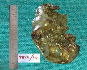
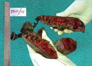
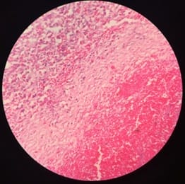
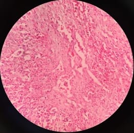
Splenic haemangioma is benign neoplasm of the spleen with fewer than 100 cases reported in the literature. They may represent small, incidental lesions that can produce significant splenomegaly and predispose to splenic rupture. These tumors are rare, usually clinically silent and diagnosed accidentally. We report a case of 65 years old male with haemangioma of spleen presenting as splenomegaly which is extremely rare and deserves a mention.

Keywords: Haemangioma, Spleen, Splenomegaly

Corresponding Author How to Cite this Article To Browse
Chandrasekhar S., Associate Professor, , Kakatiya Medical College, Warangal, Telangana, India.
Email:
Saritha C, Aneela B, Mohammed I, Chandrasekhar S, Sandhya AS. Splenic haemangioma - A rare case report. Public Health Rev Int J Public Health Res. 2016;3(1):42-47.
Available From
https://publichealth.medresearch.in/index.php/ijphr/article/view/28

Manuscript Received Review Round 1 Review Round 2 Review Round 3 Accepted
2015-12-24 2016-01-05 2016-01-15 2016-01-28
Conflict of Interest Funding Ethical Approval Plagiarism X-checker Note
No Nil Yes 9%

© 2016 by Saritha C., Aneela B., Imranali Mohammed, Chandrasekhar S., Anil S. Sandhya and Published by Siddharth Health Research and Social Welfare Society. This is an Open Access article licensed under a Creative Commons Attribution 4.0 International License https://creativecommons.org/licenses/by/4.0/ unported [CC BY 4.0].

Introduction

Splenic hemangioma is a rare disorder but remains the most common benign neoplasms of the spleen. They may represent small, incidental lesions that can produce significant splenomegaly and predispose to splenic rupture. These tumors are rare and resemble their counterparts in other organs.

The incidence of autopsy examination of their diagnosis ranges from 0.03 to 14% [1], and most often is encountered in adults between the ages of 30 and 50 [2]. Most are small lesions, usually clinically silent, diagnosed accidentally.

Sometimes, however, they may be symptomatic, manifested by splenomegaly, abdominal pain, bowel disorders, anemia and thrombocytopenia, Kasabach-Merritt syndrome (anemia, thrombocytopenia and coagulopathy) and in rare cases by spontaneous rupture of the spleen [3].

The main aim of this article is to report a rare benign pathological entity with unusual presentation as splenomegaly.

Case Report

A 65 years male presented with lump in abdomen since 6 months. He was apparently asymptomatic and suddenly developed pain abdomen. There was no history of fever. On examination, he had pallor and splenomegaly with no lymphadenopathy.

Computed tomography (plain and contrast) scan showed multiple hypodense cystic lesions of varying sizes in the splenic parenchyma extending up to the surface as described above with a large perisplenic, sub diaphragmatic haematoma – s/o ruptured splenic abscesses/Haemangioma.

Splenectomy was done and specimen was sent to the department of Pathology, MGM hospital, Warangal.

Gross Features

We received Spleen of size 14x7x3cms, weighing 275 grams. External surface was bosselated (Figure-1). Cut section showed multiple cystic spaces ranging from 0.4 cm to 2 cm in diameter filled with dark brown haemorrhagic material.(Figure-2).

public_28_01.jpg

Figure 1: Gross splenectomy specimen showing congested spleen with bosselated surface

public_28_02.jpg

Figure 2: Cut section showing multiple cystic spaces filled with dark brown haemorrhagic material.

Microscopic Features

Histologically, haematoxylin and eosin stained sections showed pools of haemorrhage with adjacent congested splenic parenchyma separated by fibrous stroma. Dilated blood filled spaces lined by single layer of bland endothelial cells, without mitosis are seen.

public_28_03.jpg


Figure 3: section showing multiplevascular spaces lined by flat endothelial cells with adjacent haemorrhagic area separated by fibrous stroma.( H& E X100)

public_28_04.jpg

Figure 4: Multiple vascular spaces lined by endothelial cells with adjacent haemorrhagic area surrounded by fibrous stroma (H&EX400)

public_28_05.jpg

Figure 5: Multiple vascular spaces lined by flat endothelial cells (H&EX40)

Discussion

Splenic hemangioma was first described by Virchow in 1863; no more than 100 cases were described in the medical literature until the year 2000 [1].

Although the etiology of splenic hemangioma is still unknown, it has been suggested that it is a congenital nevus that may or may not grow in size to become symptomatic [3,4]. Due to the slow growth of this tumor, most cases are diagnosed after the third decade of life, but cases have been described in the pediatric population in the context of various congenital syndromes like Kasabach-Merritt syndrome.

In 1945, Bostick surveyed 16 patients with splenic hemangiomas and a palpable abdominal mass and found that 62% had pain, 12% had anemia, 12% had ascites, and 18% had weight loss [4]. Another survey found that when patients presented with symptoms, they had pain, left upper quadrant fullness, or a palpable mass [1]. The most serious complication described in the literature is that of spontaneous rupture, the likelihood of which is related to tumour size [1,3].

The histology of haemangioma is characterized by the presence of benign proliferation of vascular channels that range from capillary to cavernous in size, lined by single layer of bland endothelial cells without mitosis.At immunohistochemistry studies, the capillary subtype presents with factor-VIII related antigen and CD8 negative; the diffuse subtype presents with CD68 positive; all sub- types present with CD34 positive [5,6].

Differential diagnosis to splenic haemangioma must include haemangioendothelioma [7], splenic littoral cell angioma[8], hamartoma and fibroangioma [9].

The ultrasound image shows hyperechogenic mass on spleen. Color-Doppler ultrasound allows definitive diagnosis with 84% sensitivity and 98% specificity.[10,11] The unenhanced CT-scans can show a low attenuation mass; following IV contrast, the vascular channels show centripetal fill (from the periphery inward). Larger lesions can fill more slowly and may do so incompletely and non-homogenously [12].

Splenic hemangioma may be a splenic manifestation of a systemic angiomatosis syndrome such as klippel-Trenaunay-Weber syndrome, Beckwith-Wiedemann syndrome.

Haemangiomas are not treated unless they are symptomatic or very large, with increased risk of haemorrhage; treatment is splenectomy [12]. In our case, the patient was symptomatic due to splenomegaly and splenectomy was performed for symptomatic relief.


Conclusion

Splenic hemangioma is a rare benign pathological entity with unusual presentation as splenomegaly, should be kept in differential diagnosis asit can predispose to rupture of spleen if neglected.

Reference

  1. Willcox TM, Speer RW, Schlinkert RT, Sarr MG. Hemangioma of the spleen- presentation, diagnosis, and management. J Gastrointest Surg. 2000;4(6)611-3.
  2. Hoeger PH, Helmke K, Winkler K. Chronic consumption coagulopathy due to an occult splenic haemangioma- Kasabach-Merritt syndrome. Eur J Pediatr. 1995;154(5)365-8.
  3. Husni EA. The clinical course of splenic hemangioma with emphasis on spontaneous rupture. Arch Surg. 1961;83(5)681-8.
  4. Bostick WL. Primary Splenic Neoplasms. Am J Pathol. 1945;21(6)1143-65.
  5. Arber DA, Strickler JG, Chen YY, Weiss LM. Splenic vascular tumors- a histologic, immunophenotypic, and virologic study. Am J Surg Pathol. 1997;21(7)827-35.
  6. Zukerberg LR, Kaynor BL, Silverman ML, Harris NL. Splenic hamartoma and capillary hemangioma are distinct entities- immunohisto chemical analysis of CD8 expression by endothelial cells. Hum pathol. 1991;22(12)1258-61.
  7. Budke HL, Breitfeld PP, Neiman RS. Functional hyposplenism due to a primary epithelioid hemangioendo the lioma of the spleen. Arch Pathol Lab Med. 1995;119(8)755-7.
  8. Falk S, Stutte HJ, Frizzera G. Littoral cell angioma- A novel splenic vascular lesion demonstrating histiocytic differentiation. Am J Surg Pathol. 1991;15(11)1023-33.
  1. Sencer S, Coulter-Knoff A, Day D, Foker J, Thompson T, Burke B. Splenic hemangioma with thrombocytopenia in a newborn. Pediatrics. 1987 Jun;79(6):960-6.
  2. Dubois J, Patriquin HB, Garel L, Powell J, Filiatrault D, David M, Grignon A. Soft-tissue hemangiomas in infants and children: diagnosis using Doppler sonography. AJR Am J Roentgenol. 1998;171(1)247-52.
  3. Goerg C, Schwerk WB. Color Doppler imaging of focal splenic masses. Eur J Radiol. 1994;18(3)214-9.
  4. Disler DG, Chew FS. Splenic hemangioma. AJR Am J Roentgenol. 1991;157(1)44-8.