E-ISSN:2349-4166
P-ISSN:2349-4158

Research Article

Envenomation

Public Health Review - International Journal of Public Health Research

2017 Volume 4 Number 1 January-February
Publisherwww.medresearch.in

Surgical Management of Snake Envenomation in India Current Perspective

Sheeja T M R.1*
DOI: https://doi.org/10.17511/ijphr.2017.i1.03

1* Rajan Sheeja T M, MS, DLO, MCh, DNB, Associate Professor, Department of Plastic and Reconstructive Surgery, Government Medical College, Kozhikode, Kerala, India.

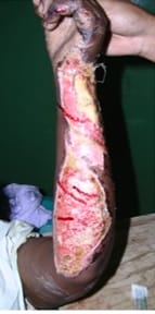
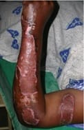
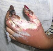
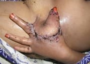
India has reported one of the highest snake bite mortality rates in the world. It is estimated that over 5 million persons per year are bitten by snakes of whom, over 1,00,000 survivors develop severe sequelae due to local wound complications. [1] The adoption of harmful first aid practices like use of tourniquets or cutting and sucking the snake bite marks further complicates wound management. National Snake Bite Protocol has been issued in 2007, with technical support from WHO to institute proper management of snake bites [9,13]. However, there is no evidence based guidelines for local wound management in snake envenomation. The local tissue problems at the bite site were effectively managed by timely administration of anti-snake venom and other supportive management in majority of patients. Cribari et al have graded the local signs of envenomation [15]. Local tissue complications are most frequently seen with bite from Viperidae. Cytotoxic enzymes in viper venom cause proteolysis, lipolysis, blisters, necrosis and gangrene. [16,18]. Surgical management of snake bite has a significant role in preventing late sequelae and permanent disability due to snake bite wounds. The aim of surgical intervention will be radical removal of all devitalized tissues, followed by reconstructive procedures using skin grafts and flaps, to minimize functional loss and maximize rehabilitation. [21] This paper explores the pathology of a snake bite wound and compiles the accepted guidelines for wound management and also the do’s and don’ts pertaining to the bite areas following a snake bite.

Keywords: Debridement, Envenomation, Fasciotomies, Snake venom, Surgical management, Wound coverage

Corresponding Author How to Cite this Article To Browse
Rajan Sheeja T M, MS, DLO, MCh, DNB, Associate Professor, Department of Plastic and Reconstructive Surgery, Government Medical College, Kozhikode, Kerala, India.
Email:
Sheeja T M R. Surgical Management of Snake Envenomation in India Current Perspective. Public Health Rev Int J Public Health Res. 2017;4(1):13-19.
Available From
https://publichealth.medresearch.in/index.php/ijphr/article/view/58

Manuscript Received Review Round 1 Review Round 2 Review Round 3 Accepted
2017-02-06 2017-02-14 2017-02-20 2017-02-27
Conflict of Interest Funding Ethical Approval Plagiarism X-checker Note
No Nil Yes 8%

© 2017 by Rajan Sheeja T M and Published by Siddharth Health Research and Social Welfare Society. This is an Open Access article licensed under a Creative Commons Attribution 4.0 International License https://creativecommons.org/licenses/by/4.0/ unported [CC BY 4.0].

Introduction

India has reported the highest snake bite mortality rates in the world. It is estimated that over 5 million persons per year are bitten by snakes of whom 1,00,000 develop severe sequelae.[1] In India, WHO estimate the magnitude of this problem to be 83,000 bites per annum with 11,000 deaths [2,3].

India’s first national survey on the cause of death, called Million Death Study (2001-2003) by Registrar General of India and the Centre for Global Health Research reports an estimate of 48,000 annual deaths by snake bites[4]. Most of the fatalities are due to failure of reaching a health facility with provisions for definitive treatment.

Compounding to this problem are the adoption of harmful first aid practices for snake bite wound management and interventions by traditional healers.

WHO considers snake bite to be a neglected disease and WHO/SEARO has published guidelines for clinical management of snake bite in south east Asian regions [5].

Indian States with the high incidence of snake bite cases are Tamil Nadu, West Bengal, Uttar Pradesh, Maharashtra and Kerala.[6,7,8] Kerala is considered to have higher standards of public health care system when compared to the rest of India. The 5 year statistics of a dedicated snake bite unit from a tertiary care teaching hospital in North Kerala is depicted in Table 1.

There were a total of 1.77% (n=28) deaths among the poisonous bites and only 0.1% (n=10) local wound problems required surgical consultation. The local tissue problems at the bite site were effectively managed by systemic anti-snake venom administration and other supportive management in majority of patients.


Table 1: 5 year statistics of a dedicated snake bite unit from a tertiary care teaching hospital in Kerala State

Year Poisonous bite Nonpoisonous bite Death Surgical intervention
Male Female Total Male Female Total
2012 222 81 303 1822 397 2219 10(3.3%) 1
2013 270 129 399 1106 424 1530 9(2.3%) 2
2014 263 100 363 1164 489 1653 1 (0.3%) 2
2015 206 95 301 1087 617 1704 2 (0.66%) 3
2016 139 72 211 1044 594 1638 6 (2.84%) 2
Total 1577 8244 28 10

There is no evidence based guidelines for local wound management in snake envenomation. This paper explores the pathology of a snake bite wound and compiles the accepted guidelines for wound management and also the do’s and don’ts pertaining to the bite areas following a snake bite.

Snakes populate all continents of the world with the exception of the Artic, Antarctic and a few small islands1. In India, more than 200 species of snakes are identified, 52 of which are poisonous. The three major families of venomous snakes are the Elapidae (Cobra, King Cobra, Krait, Coral Snake), Viperidae (Russel viper, Saw scaled viper, Hump nose viper, Russell’s viper of Croatalinae subfamily) and the Hydrophidae (sea snakes).

In India, the “big four” - the Krait (Bungarus caerulens/fasiatus/sindanus), Indian cobra (Naja Naja/kaouthia/oxiana), Russels viper (Daboia russelii) and Saw scaled viper (Echis carinatus

/sochureki) and the most common poisonous snakes.[9,10] But among the four, two thirds of bites are due to the Saw scaled viper, a fourth to Russel’s viper and only a small proportion of bites to Cobra and Kraits [2,10].

Snakes are equipped with a venom apparatus in order to procure the prey and help in digestion. The toxic component of snake venom includes enzymes, polypeptides, glycoproteins and several low molecular weight compounds [11].

The chemical effects of the components and the inflammatory response elicited result in the toxic effects of snake venom in the victim’s body. Table 2 shows the main components of snake venom and their toxic effects in the body. Salient features of the Big 4 snake bites in India are shown in Table 3 [12,13,14].


Table 2: The main components of snake venom and their toxic effects in the body

Snake venom components Toxic effects
Proteolytics
Enzymes – Hydrolases, Metalloproteases, Hyaluronidase
Oedema, Blistering,
Necrosis
Spread toxins in tissue planes
Haemorrhagins Damage vascular endothelium
Phospholipase A2 Cytochrome c level mitochondrial inhibition, cell damage
Neurotoxins
-Alpha polysynaptic (Cobra)
-Presynaptic (Krait)

Binds to motor end plates
Release and deplete Acetyl choline
Others – Peptides, Nucleotides, Amines, metals, Lipids Systemic toxicity
Procoagulants Activate and deplete clotting à consumption coagulopathies (Viperidae & some Elapidae)
Haemorrhagins Zinc Metalloproteinases – damage Vascular endothelium and cause bleeding.

Table 3 : Salient clinical features of the “Big 4” snake bites in India

Cobra Krait Russel’s viper Saw scaled viper
Neurotoxic
Marked local pain & swelling
Ecchymosis, Necrosis
No coagulopathy
Neurotoxic
Not much local signs
Small undiscernible puncture marks
Hemotoxic
Coagulopathy
Marked local swelling
Early renal failure
Mild neurotoxicity
Hemotoxic
Marked local swelling, discoloration, coagulopathy,
No neurotoxicity

Severity of envenomation depends on several factors. Fang marks with deep penetration of skin, multiple strikes or bites by a younger the snake indicate more venomous bites. Generally, 50% Russel viper bites, 30% cobra bites and 5-10% saw scaled viper bites are non-venomous “dry bites”[4,9].

When the venom has been injected, there will be local symptoms and signs like fang marks with progressive increase in local pain, swelling with proximal extension, bleeding from puncture wounds, bruising, lymphangitis and tender painful regional lymphadenopathy [1,5]. There will be subsequent blistering of inflamed skin with local infections and abscess formation.

In late cases, areas of necrosis with demarcation, loss of sensation and putrefaction will set in.

There will be remarkably less local symptoms and signs on bites by common Krait and sea snakes. Faulty practices in snake bite wound management can cause misleading clinical features without envenomation. Constricting tourniquets cause excessive local pain, swelling and congestion.

Traditional practices of incising into fang marks and attempts to suction venom, burning the area over flame, cryotherapy or application of herbal remedies will cause inflammation, blistering and infection leading to diagnostic dilemma [9,12]. The long term sequelae of improperly treated snake bite wounds include chronic ulceration, infection, osteomyelitis, joint contractures, arthritis and marjolin’s ulcers over non healing wound.

National Snake Bite Protocol has been issued in 2007, with technical support from WHO to institute proper management of snake bites [9,13]. This document emphasizes on preventive measures against snake bite and the need for transporting victim to nearest treatment facility within the shortest possible time.

The first aid that is currently recommended to be administered by self or community volunteers is based on the mnemonic- “Do it R.I.G.H.T”. [R = Reassure, I = Immobilize, GH = Get to Hospital Immediately, T = Tell the doctor the systemic symptoms that have appeared during transport]

The protocol states that certain traditional methods are to be discarded since they are found to do more harm than good. The practices to be avoided are as follows:

  • Cutting, puncturing, burning, sucking the wound or applying boiling water, electric shock, ice packs or chemicals over wound are to be strictly avoided because they are ineffective in removal of venom and increases wound complications [14].
  • Washing snake bite wound with soap and water is to be avoided since this can lead to increase in venom spread by stimulating the lymphatic system.
  • Tourniquets are now contraindicated. The traditional practice of tying a tight tourniquet carries the risk of ischemia and loss of limb and increases the incidence of necrosis in poisonous bites. In addition, they may lead to embolism and shock in viper bites and sudden neurotoxic blockade during tourniquet release.

Most importantly, they do not work and have been experimentally shown not to slow venom spread. Furthermore, the false sense of security given by the tourniquet can inadvertently delay the transport of patients to hospital.

  • Pressure Immobilization Method (PIM) are inadvisable.

PIM was developed by Sutherland in 1974 in Australia for viper bites. This involves bandaging of the bitten limb with crepe bandage and integral splint for full immobilization. Recommended pressures are of 40-70mm Hg in the upper limb and 55-70mm Hg in the lower limb [5]. The PIM is not currently recommended for use in Indian rural context due to technical reasons9.

Once the victim reaches hospital, the snake bite treatment protocol will be directed initially for patient assessment at presentation and to provide basic resuscitation if needed [1,2,4,5]. If there is evidence of skin puncture. broad spectrum antibiotic and tetanus prophylaxis are administered.

Pain management is done with Paracetamol, and sometimes with Tramadol. NSAIDs are contraindicated since they can cause bleeding. Tourniquets are to be removed with care. If they are occluding distal pulse, a BP Cuff is applied proximally before removal.

In the diagnostic phase, all patients with suspected snake bite are to be kept under observation for a minimum of 24 hours, during which the general signs for Viperidae or Elapidae envenomation will be periodically assessed. The most reliable coagulation test carried out at bedside in most centers is the 20-minute whole blood clotting test (20 WBCT) [1,5,9].

Other useful tests include hematocrit, platelet count, coagulation profile, FDP and D dimers, peripheral smear, urine test for RBC, proteins, myoglobin, hemoglobin, renal parameters, O2 saturation, postural blood pressure, ECG, USG and ABG [1] (which maybe done if facilities are available). It will take some time before ELISA for identification of species of snake and level of envenomation becomes available for general management of all.

ASV will be administered only if there is evidence to suggest systemic envenomation by coagulopathy or neurotoxicity or if there are features of severe current local envenomation.

ASV administration can be associated with anaphylactic reactions in the acute phase and serum sickness in later stages. The center should be well equipped on prophylaxis of the inadvertent reactions before ASV administration. There is no role for local administration of ASV near bite site since it is ineffective, painful and increased intra compartment tension1

Features of severe current local envenomation as per National snakebite management protocol are severe current local swelling involving more than half of the bitten limb (in the absence of tourniquet), severe swelling after bites on the digit (toes & fingers), bite from a known necrotic species, rapid extension of swelling – beyond wrist and ankle within few hours of bite on hands / feet, swelling continues to progress one hour after the removal of tourniquet.[9] Cribari et al have graded the local signs of envenomation as given in Table 4.[15].

Table 4: Grades of local envenomation Chris Cribari 2004

Grade 0 Fang marks, swelling and erythema around fang mark less than 2.5cm, pain & tenderness are minimal, no systemic symptoms
Grade 1 Fang marks, immediate pain with the bite, swelling & erythema
Grade 2 Fang marks, immediate severe pain, swelling & erythema 15-40cm, mild systemic symptoms, abnormal lab findings
Grade 3 Fang marks, immediate severe pain, swelling and erythema more than40cm petechiae & bullae, modular systemic symptoms Bleeding and/or DIC Abnormal lab values
Grade 4 Fang marks, signs of multiple envenomation sites, immediate severe pain, severe systemic signs like coma, shock, bleeding, DIC & paralysis

Surgical management in Snake bite wounds:

Surgical intervention is needed for:

  1. Debridement of necrotic tissues
  2. Fasciotomies – in compartment syndrome
  • Wound coverage

If the victim receives timely management with resuscitation and ASV administration, it can significantly reduce the local swellings and complications, curbing the need for surgical management. Most often, surgical intervention will be needed in late presentations or if the degree of envenomation was high.

Surgery can be undertaken only after the patient has been stabilized, ASV protocol completed and coagulation profile restored. Sometimes the extent of deeper tissue damage may be more than the appearance around fang marks.


Small blisters can be left undisturbed. Larger bullae can be aspirated if tense and spreading [11,16.]. The local swelling can mislead us to the erroneous diagnosis of compartment syndrome. There is little objective evidence that intra-compartmental pressure ever reaches prescribed limit of more than 30mm Hg to warrant a fasciotomy in the Indian context of snake bite wounds. Visual impression is a highly unreliable guide for ICP assessment [17,18].

Fasciotomy does not remove or reduce envenomation and should be undertaken with care, only after restoration of coagulation status. Unnecessary fasciotomies can cause profuse bleed, residual skin loss, expose tendons and neurovascular bundles and devitalize them, cause hematomas, persistent oozing and shock.

Local tissue complications are most frequently seen with bite from Viperidae. Cytotoxic enzymes in viper venom cause proteolysis, lipolysis, blisters, necrosis and gangrene. [16,18] This will be further complicated by hematological, hemotoxic and myotoxic complications. Since dorsum of hand feet lack abundant soft tissue covering, the tendon, nerves, vessels and joints are prone to injury and exposure following loss of necrosed skin [18,19,20].

The aim of surgical intervention for wound coverage will be radical removal of all devitalized tissue, including amputations, followed by reconstruction procedures to minimize, functional loss and maximize rehabilitation [21]. Full or split thickness skin grafts (fig 1 & 2), cross finger flaps, thenar flaps, groin flap (fig 3,4 &5) or hypogastric flaps are the most frequently used reconstructive option for hand [18]. Leg defects can be covered with appropriate loco regional flaps.

VAC dressings can be used prior to grafting. There is insufficient evidence for microvascular free flap surgeries in the context of snake bite wounds. If used, additional precautions will be needed since Heparin or Aspirin used as anticoagulants after microvascular anastomosis are contraindicated in snake bite patients [9]. In our series of patients only 0.1% of total envenomation required surgical intervention which shows the effectiveness of early ASV protocol in curbing the local tissue damage effects of snake venom.

Still it is important to remember that though surgery is not as important as ASV treatment, there should be no delay in timely referral and interventions to ensure maximum functional restoration especially of hands [17,18,20,21].

Surgical management of snake bite has a significant role in preventing late sequelae and permanent disability due to snake bite wounds.

Huang TT et al have used excisional treatment in the management of snake bite based upon the finding that bulk of deposited venom will remain in the area of bite and much removal of the tissue containing injected venom can eliminate the local tissue toxicity and reduce the magnitude of systemic toxicity. There was a marginal increase in skin flap necrosis followed the excision and may not be suitable in the Indian context [22].

The limited efficacy of anti-snake venoms on local tissue damage is not due to the lack of neutralizing antibodies in the anti-venom, but rather due to the rapid development of local pathology before that makes it difficult to access the area of insensible damage.

The future directives of management of snake envenomation is focusing on the development of drugs that can inhibit the hydrolyzing enzymes like metallo-protein phospholipase A2 and hyaluronidase which are responsible for the extensive tissue damage [23,24].

The use of such drugs can be expected to obviate the need for high risk surgical interventions in the coming days. In the mean while community education and ensuring their participation can reduce the morbidity and mortality of snake bite envenomation considerably in the Indian context.

Acknowledgement

The author wishes to duly acknowledge the staff of Department of General medicine, Government Medical College, Kozhikode for their contributions in the preparation of this article.

Reference

  1. Ahmed SM, Ahmed M, Pal J. Emergency treatment of a snake bite- Pearls from literature. J Emerg Trauma Shock. 2008 Jul-Dec;1(2)97-105.
  2. Singh S, Singh G. Snake Bite- Indian Guidelines Protocol. Toxicology. 2013.

  1. Kasturiratne A, Wickramsinghe AR, DeSilva N, et al. Global burden of snakebite- A literature analysis and modelling based on regional estimates of envenoming and death. PLOS Med. 2008;5;218.
  2. Anghore D, Sharma A, Singh S, et al. Treatment of snake bite in India- A review. IJPTP. 2015;6(4)2635-2641.
  3. David A Warrel. Guidelines for management of snake bites. WHO Regional Office for South-East Asia. 2010.
  4. Hati AK, Mandal M, De MK, et al. Epidemiology of snake bites in the district of Burdwan, West Bengal. J Indian Med Assoc. 1992;90;145-147.
  5. Gaitonde BB, Bhattacharya S. An epidemiological survey of snake-bite cases in India. Snake. 1980;12;129–33.
  6. National Snake bite Management Protocol. Ministry of Health and Family Welfare. Govt of India, 2009 With Technical Support from:
  7. Punde D P. Management of snake bites in rural Maharashtra- A 10-year experience. Nat Med Journal of India. 2005;18(2)71-75.
  8. Saini RK, Sharma S, Sing S, Pathania NS. Snake Bite Poisoning- A preliminary report. J Assoc Phys India. 1984;39;195.
  9. Toschlog EA, Bauer CR, Hall EL, et al. Surgical consideration in the management of pit viper snake envenomation. J Am Coll Surg. 2013 Oct;217(4)726-735.
  10. Ghosh S, Mukhopadhyay P, Chatterjee T. Management of snake bite in India. Journal of Assoc Physicians of India. 2016 Aug;64;11-14.
  11. Jobin A, Rai N. New aspects for treatment of snake bite- a review. IRJP. 2012;3(11)1-15.
  1. Simpson ID, Norris RL. Snakes of Medial Importance in India, Is the concept of the “Big 4” Still relevant and useful?. Wilderness Environ Med. 2007;18;2-9.
  2. Cribari C. Management of poisonous snake bites. American College of Surgeons Committee on Trauma. 2004. Available at:
  3. Kose R. The management of snake envenomation, Evaluation of 21 snake bite cases. Turkish Journal of Trauma, Emergency Surgery. 2007;13;307-12.
  4. Mabvuure NT, Malahias M, Hindocha S, et al. Acute compartment syndrome of the limbs, Current-concepts and management. Open Orthop J. 2012;6(9)535-543.
  5. Borzkut M, Kulahi Y, Zon F, Kapi E. The management of Pit Viper Envenomation of the Hand. HAND. 2008;3;324-33.
  6. Malahias M, Jordan D, Hughes O, et al. Bite injuries in the Hand- Microbiology, Virology and Management. Open Orthop J. 2014;8(1)157-6.
  7. Gras S, Ptantefeve G, Band F, Crippaux JA. Snake bite on the hand- Lessons from two clinical cases illustrating difficulties in surgical indication. J Venom Anim Toxins incl Trop Dis. 2012;18(4)467-477.
  8. Watt CH Jr. Treatment of poisonous snake bite with emphasis on digital dermotomy. South Med. 1985;78;694-9.
  9. Huang TT, Lynch JB, Duane L et al. The use of excisional therapy in the management of snakebite. Ann Surg. 1971;May;179(5)598-605.
  10. Jose Marie Gutierroz, RDG Theakston, Warrel DA. Confronting the neglected problem of snake bite envenoming- The need for a global partnership. PLOS Media. 2006 June;3(6)727-31.

  1. Rucavado A, Escalante T, Guitierrez JM. Effect of metalloproteinase inhibition batimastat in the systemic toxicity induced by Both rops as per snake venom understanding the role of Metalloproteinase in envenomation. Toxicon. 2004;43;41.

Figure Legends

Figure 1: Snake bite raw area left forearm

public_58_01.jpg

Figure 2 : Wound coverage with Split thickness Skin graft

public_58_02.jpg

Figure 3 : Snake bite wound left hand with tissue necrosis

public_58_03.jpg

Figure 4 : Wound covered with pedicled Groin flap after debridement

public_58_04.jpg

Figure 5 : Wound covered with Groin flap

public_58_05.jpg